The right dental X-ray depends on the question your dentist needs to answer: decay between teeth calls for a different view from a painful root, an impacted wisdom tooth, or an implant site. By matching symptoms and treatment plans to each scan, you can ask for the smallest useful image and avoid treating “digital” as if it were a specific X-ray type.
Key takeaways
- Choose the scan based on the suspected problem, not a routine package.
- Bitewing X-rays reveal cavities between teeth; periapical images show roots and bone.
- Panoramic X-rays show both jaws, while CBCT provides three-dimensional detail.
- Ask which clinical question the scan will answer before having dental imaging done.
Start with the dental problem, not a routine X-ray package
The answer to “what type of dental x ray do i need in dombivli” follows the suspected problem, not a routine package. Your dentist considers the tooth’s location, symptoms, age, caries or gum risk, planned treatment, and any recent usable images before prescribing an image.
- Start with the examination. A visual check, symptom history, pulp testing, and periodontal measurements can identify some causes of pain or gum disease without imaging.
- Take an image when the suspected problem lies below the gum, between teeth, inside a tooth, around a root, or in bone, or when treatment such as root-canal therapy, extraction, implants, or orthodontics needs information the examination cannot provide.
- Review recent images first. Repeat them only when they are not diagnostically adequate, do not cover the needed area, or cannot answer the current clinical question.
A normal radiograph does not rule out early decay, a crack, pulp inflammation, some periodontal problems, or soft-tissue causes of pain. A tooth can hurt before visible bone changes appear, so a normal image does not end the diagnosis.
The smallest useful image is usually preferred. An intraoral view may answer a focused question, while a larger scan is reserved for a broader area or a specific treatment decision. Ask what question the proposed X-ray will answer and how its result will change your care.
Match each X-ray type to what it can actually show
The right image depends on the anatomy your dentist needs to see, not on whether the machine is labelled digital. Each X-ray answers a different clinical question.
| Type | What it shows | When it fits |
|---|---|---|
| Bitewing | Crowns of back teeth and nearby crestal bone, not full roots | Decay between teeth and crestal bone height |
| Periapical intraoral | One complete tooth, root tip, and surrounding bone | Toothache, infection, trauma, root resorption, or root-canal assessment |
| Occlusal | A larger section of the upper or lower jaw | Finding teeth, stones, or lesions across a wider area |
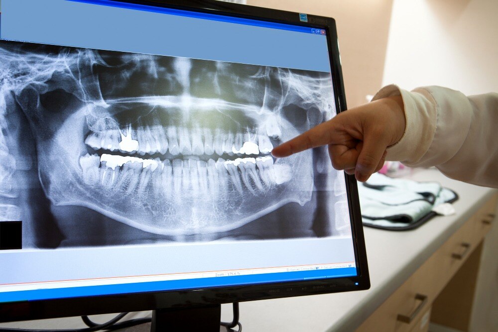
| Panoramic | Both jaws, wisdom teeth, developing teeth, and a broad view of the jaw-joint regions | Wisdom teeth, multiple unerupted teeth, fractures, lesions, or broad planning |
| Lateral cephalometric | Jaw relationships, tooth inclination, facial growth, and treatment changes | Orthodontic planning |
| CBCT | Three-dimensional implant anatomy, nerve relationships, complex roots, fractures, or lesions | When two-dimensional images cannot answer a specific question |
A bitewing gives finer detail for early decay between back teeth than a panoramic image. Panoramic imaging covers more anatomy, but magnification, overlap, and geometric distortion reduce detail.
CBCT is not an automatic upgrade for a routine examination, extraction, or implant. Its larger scan volume brings greater exposure, artefacts, and cost; use the smallest field that answers the question.
If you search for an oral X ray in Dombivli, ask which anatomy the proposed image covers and what decision it will change.
Use your symptoms and treatment plan as the decision guide
Food trapping or suspected decay between teeth usually calls for bitewings. A focused toothache, swelling, trauma, root-canal assessment, root resorption, or bone loss around one tooth usually calls for a periapical view.
| Likely problem or treatment | Usually suitable image | What it answers |
|---|---|---|
| Food trapping or decay between back teeth | Bitewing | Shows crowns and nearby crestal bone, not full roots |
| Toothache, swelling, trauma, root-canal review, root resorption | Periapical | Shows one complete tooth, root tip, and surrounding bone |
| Wisdom teeth, multiple missing or unerupted teeth, broad jaw assessment, suspected fracture | Panoramic | Shows both jaws, jaw-joint regions, and developing or impacted teeth |
| Orthodontic planning | Lateral cephalometric | Shows jaw relationships, tooth inclination, and facial growth |
| Implant planning, difficult impaction, complex root-canal anatomy, suspected root fracture, or jaw lesion | CBCT | Provides three-dimensional detail when two-dimensional images cannot answer the question |
Dentures and full-mouth rehabilitation often need several views, chosen around the remaining teeth and supporting bone. Ordering every view at once adds exposure, cost, and review time without improving the plan.
Jaw-joint concerns can begin with an examination and panoramic image. Advanced imaging follows only when those findings and your symptoms leave a specific question unanswered. When comparing dental imaging in Dombivli, ask which clinical question the proposed image will resolve rather than choosing a routine package.
Know what happens during the scan and how to prepare
Expect the setup to take longer than the exposure itself. For an intraoral X-ray, the operator places a small sensor inside your mouth beside the tooth and asks you to hold it by biting gently. Stay still until the exposure ends.
For a panoramic or lateral cephalometric scan, you stand or sit with your head against supports while the machine aligns your face. A CBCT scan also requires stillness as the scanner rotates around your head.
Retakes happen when you move, bite on the sensor incorrectly, or the image misses the tooth, root, jaw area, or other anatomy needed for the diagnosis. Follow these preparation steps:
- Eat and drink normally unless the imaging provider gives different instructions.
- Remove removable jewellery, glasses, hair accessories, and dentures when asked. Metal can obstruct parts of the image.
- Tell the dentist or operator if you are pregnant or could be pregnant, and report relevant medical history, recent trauma, and any recent dental images or reports.
- For a child, expect child-appropriate exposure settings and careful positioning. A smaller patient does not automatically need the same set of images as an adult.
- Use the smallest scan that answers the clinical question. A focused intraoral image avoids covering unnecessary anatomy; CBCT is reserved for questions that two-dimensional images cannot answer.
Ask the Dombivli provider what clinical question the scan answers
A dental X-ray clinic in Dombivli should explain the clinical question before recommending an image. Ask which modality is proposed, what anatomy it covers, and how the result could change your diagnosis or treatment plan.
| Modality | Coverage | Clinical question |
|---|---|---|
| Intraoral image | One or a few teeth, roots, and nearby bone | Decay, pain, infection, or root-canal assessment |
| Panoramic image | Both jaws, wisdom teeth, developing teeth, and jaw joints | Broad assessment of teeth, eruption, or jaw problems |
| Cephalometric image | Side view of the skull, jaws, and facial profile | Orthodontic growth, jaw relationships, and tooth inclination |
| CBCT | Three-dimensional section of the teeth and jaws | Implant anatomy, impacted teeth near nerves, complex canals, root fractures, or lesions |
Before booking, ask:
- Could a smaller CBCT field of view, or a single intraoral image, answer the question instead?
- Will the dentist review recent, diagnostically adequate images before ordering another scan?
- If a repeat is needed, what was missing, unclear, or changed?
- What treatment decision will the new image affect?
- Can you share previous reports or image files?
“Digital” describes image capture or storage. It does not prove wider coverage, accuracy by itself, or clinical necessity. Dr. Sunil Shahane Dental Clinic is relevant when the dentist connects your examination, symptoms, and treatment plan to the imaging choice instead of offering every patient the same panoramic or full-mouth package.
Related service
Digital Dental X-ray At the clinic of Dr. Pooja Ganu-Agnihotri in Dombivli, Digital Dental X-ray technology provides fast, accurate, and comfortable diagnostic imaging... View service → |
Frequently asked questions
How do I know what type of dental X-ray I need in Dombivli?
Your dentist chooses based on the tooth location, symptoms, suspected decay or gum disease, age, and planned treatment. Existing usable images can prevent unnecessary repeat scans.
What can different dental X-rays show?
Bitewing X-rays show decay between teeth. Periapical X-rays show the full tooth, root, and surrounding bone. Panoramic X-rays show both jaws and all teeth in one image. CBCT creates three-dimensional views for complex implant, surgical, orthodontic, or jaw assessment.
How should I prepare for a dental X-ray?
Tell the provider if you are pregnant or might be pregnant, remove glasses and metal objects near your head, and follow instructions about positioning. Most dental X-rays need no fasting.
What should I ask a dental X-ray clinic in Dombivli?
Ask what clinical question the scan answers, why that type is needed, whether a recent image is usable, how the scan will affect treatment, and when you will receive the report or images.
Keywords